|
Unilateral and Bilateral Locked Facets
General Considerations
- Normal relationships of facet joints: Inferior articulating facet of body above (blue arrow) lies posterior to superior facet of body below (red arrow)

Ligamentous Injuries
- Mechanism is flexion/distraction
- Injury is to the posterior spinal ligamentous complex
- Unstable in flexion; stable in extension
- If unrecognized, can lead to progressive neurologic damage
- Imaging Findings
- Widening of the interspinous distance
- Slight anterior subluxation of one vertebral body on another
- Widening of the facet joint
- Usually the posterior aspect
- Disk space narrower anteriorly than posteriorly
- Sharp angle kyphosis

From Seminars in Roentgenology, Jan 1978 John H. Harris, Jr.
Posterior ligamentous structures involved in flexion injury
are (a)supraspinous ligament (b) interspinous ligament (c) facet joint capsule (d) posterior longitudinal ligament
- Degrees of ligamentous injury
- Subluxation of vertebral body
- Perched facet
- Locked facets
Unilateral Locked Facet
- Mechanism is flexion/distraction and rotation
- Only 30% associated with neurologic defect
- Most often occurs at C4-5 and C5-6
- Inferior articular facet of superior vertebral body is locked in front of the superior facet of the more inferior vertebral body but only on one side
- Imaging Findings
- Subtle
- Slight anterior subluxation of one vertebral body on the one below
- Usually less than 25% of the width
- On lateral view of cervical spine, some bodies appear true lateral below level of injury and oblique above level of injury
- Spinous processes do not align on frontal film
- Spinous processes of inferior vertebrae displaced toward the locked side
Bilateral Facet Dislocation
- Severe flexion injury
- Both anterior and posterior ligamentous structures are disrupted at site of injury
- More superior vertebra subluxes forward by 50% or more of the body below
- Usually occurs in lower cervical spine

Locked facets. Body of C4 is subluxed anteriorly on C5.
The inferior facet of C4(blue arrow) lies anterior to the superior facet of C5 (red arrow) below it.
The normal relationship has the inferior facet posterior to the superior fact below it.
- May have associated fractures of the laminae and vertebral arch
- Quadriplegia frequently develops
- If there is a fracture through posterior elements, less chance of neurologic injury as cord can decompress
- 85% neurologic deficits with locked facets


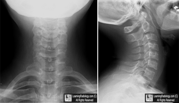
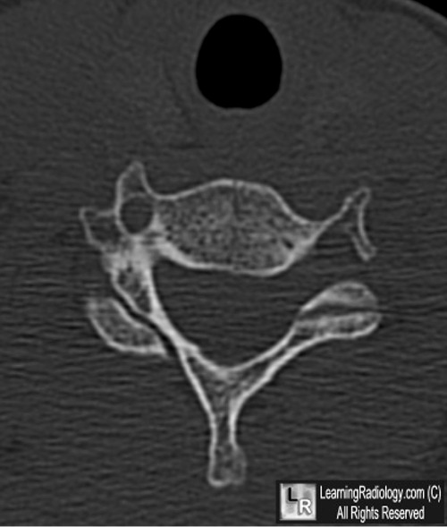
Unilateral Locked Facet. Top. On frontal view of cervical spine, at level of the locked facet (C5), the spinous process is displaced towards the right (black arrow), out of alignment with those below it (white arrows). The lateral view demonstrates the "bow-tie" sign at the level of the locked facet. The superior facet of C6 (yellow arrow) is posterior to the inferior facet of C5 (blue arrow).Bottom: Axial CT at the level of C5 shows the normal facet relationship on the left (hamburger on a bun) (white arrow) while the locked facet is seen on the right (reverse hamburger on a bun) (yellow arrow).
For these same photos without the arrows, click here and here
For more information, click on the link if you see this icon 
Seminars in Roentgenology, Jan 1978
Emergency Medicine Clinics of North America, August 1965
|
